What the latest evidence says about the conditions that keep patients off their feet — and how modern treatment is changing outcomes
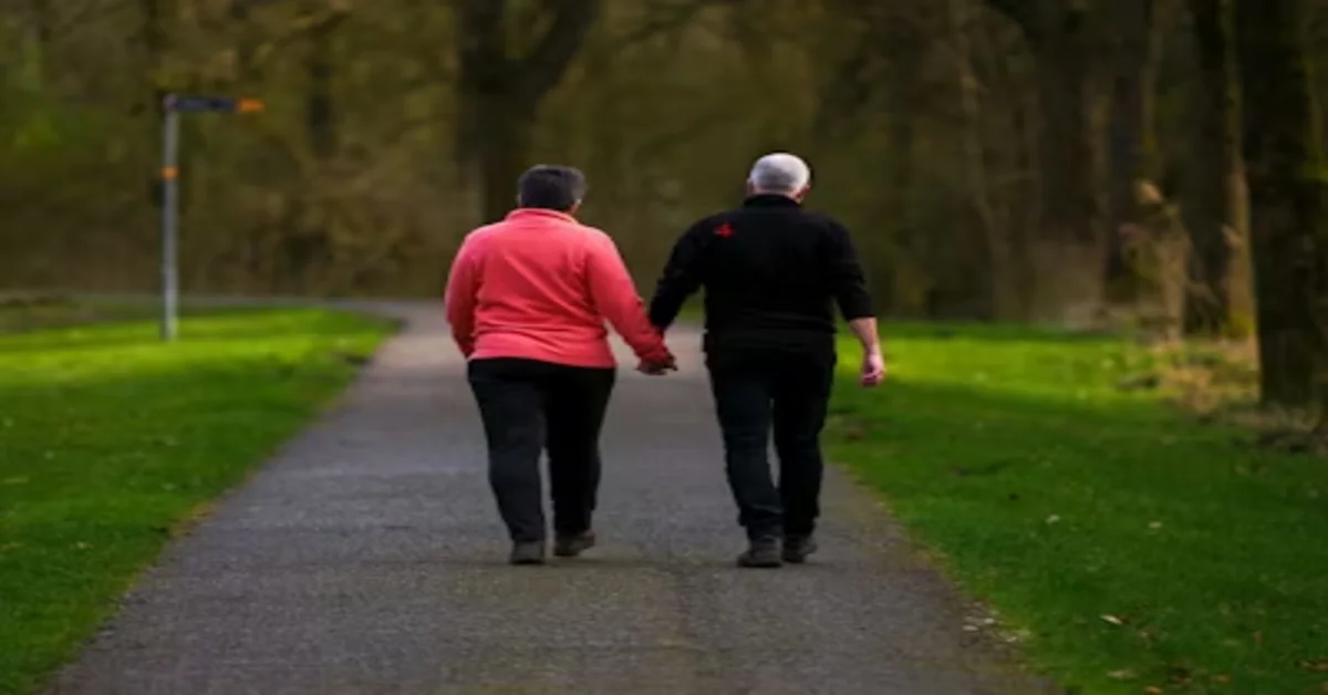
There is a particular cruelty to heel and tendon pain. It does not announce itself with a dramatic injury. It tends to arrive quietly — a tightness in the first few steps out of bed, an ache that fades after a warm-up and returns with a vengeance the next morning, a stiffness in the back of the ankle that was tolerable last month and is limiting this one. By the time most patients seek care, they have been adapting their gait, their footwear and their activities for far longer than they should.
Posterior heel and arch conditions — Achilles tendinopathy in its various forms, plantar fasciitis, and the bony prominence at the back of the heel known as a Haglund’s deformity or heel bump — are among the most common reasons adults present to foot and ankle specialists. They are also among the most treatable, provided the right intervention is matched to the right diagnosis at the right time.
This article works through each condition in turn: what is actually happening in the tissue, where treatment has historically fallen short, and what newer approaches are producing better outcomes for patients who have not responded to first-line care.
Achilles tendinopathy treatment: why the old approach often failed
Achilles tendinopathy is not a single condition. It is a spectrum of tendon pathology driven by overload — a cumulative failure of the tendon’s capacity to adapt to the demands placed on it. The terminology has evolved deliberately over the past two decades: we no longer call it tendinitis, because histological studies showed that classical inflammatory cells are largely absent in chronic cases. The tissue changes are degenerative, not inflammatory in the traditional sense, which is why anti-inflammatory medication alone does so little for most patients.
The mid-portion and the insertion are the two anatomically distinct presentations, and they respond to treatment differently — a point that is still frequently missed in primary care.
Mid-portion Achilles tendinopathy treatment is now anchored in progressive tendon loading, based on the work of researchers including Hakan Alfredson and subsequently Jill Cook and Craig Purdam. Heavy slow resistance (HSR) protocols — involving slow, loaded calf raises performed on both flat and inclined surfaces — have become the gold standard first-line intervention. When executed consistently over 12 weeks, they produce clinically meaningful improvements in pain and function in the majority of patients. The evidence base here is among the strongest in musculoskeletal physiotherapy.
For patients who do not achieve sufficient improvement through loading alone, adjunct therapies have expanded the toolkit. Extracorporeal shockwave therapy (ESWT) has a well-supported evidence base for both mid-portion and insertional Achilles tendinopathy treatment, stimulating neovascularisation and promoting collagen remodelling in tissue that has become dysregulated. PRP (platelet-rich plasma) injections are increasingly used as an adjunct in recalcitrant cases, though the evidence remains more heterogeneous than for shockwave. GTN patches, once widely used, have largely fallen out of favour following updated trial data.
The key word in all of this is “recalcitrant.” Most patients with mid-portion Achilles tendinopathy who receive a proper loading programme early, with adequate guidance and progression, do very well without injections or procedures. The problem is that most patients do not receive a proper loading programme early. They receive rest advice, a cortisone injection, and a generic physiotherapy referral. Cortisone is now contraindicated in most tendinopathy guidelines because it weakens tendon structure without addressing the pathology — and in the Achilles, it carries a risk of rupture that should not be taken lightly.
Achilles tendon rupture treatment: the case for non-surgical management has strengthened
A complete Achilles tendon rupture is a different and more serious injury than tendinopathy. It typically occurs during a sudden explosive push-off — a sprint start, a jump, a sudden change of direction — and is often described by patients as feeling like they have been kicked or struck from behind when nobody was near them.
The debate between surgical and non-surgical Achilles tendon rupture treatment has shifted considerably over the past ten years. Randomised controlled trials — most notably the UK UKSTAR trial — have demonstrated that functional rehabilitation in a controlled equinus cast or boot, with early weight-bearing and progressive range-of-motion exercises, produces outcomes equivalent to surgical repair in terms of re-rupture rate, functional recovery and return to activity. For most patients, particularly those who are not elite athletes and whose rupture is managed promptly, the non-surgical pathway avoids surgical risk without sacrificing outcome.
Surgical repair remains appropriate for certain presentations — athletes with high functional demands, cases where the tendon ends are significantly gapped and non-surgical approximation is inadequate, or patients presenting late. The critical factor in either pathway is the quality and adherence to the rehabilitation programme, which determines functional outcomes more than the initial treatment choice.
Plantar fasciitis treatment: what actually works in the medium and long term
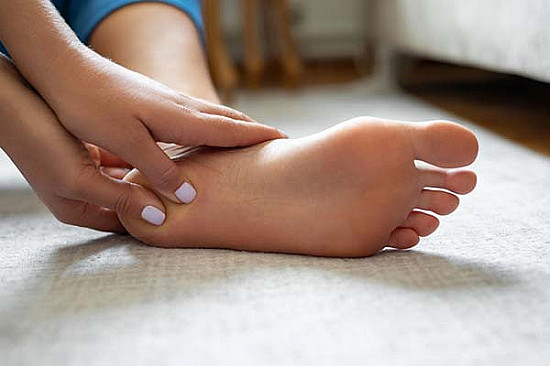
Plantar fasciitis is the most common cause of heel pain in adults, affecting roughly one in ten people at some point in their lives. The classic presentation — sharp, stabbing pain in the heel on the first steps of the morning, improving after five to ten minutes of walking and returning with prolonged standing or activity — is familiar to most clinicians and to most of the patients who walk through the door.
The evidence on plantar fasciitis treatment is unusually well developed for a musculoskeletal condition. We know that most cases — approximately 90% — resolve within 12 months with conservative management. We also know that for the 10% who do not resolve, and for the majority of patients who want resolution faster than 12 months, there are effective interventions. A comprehensive overview of current plantar fasciitis treatments covers the full treatment ladder in detail.
The evidence-supported first-line interventions are stretching (particularly plantar fascia-specific stretching, performed seated with the toes extended before the first step of the morning), custom or prefabricated orthotics with good arch support, calf flexibility work, and load management. Night splints — which maintain the fascia in a lengthened position overnight, reducing morning stiffness — have a solid evidence base and are underused.
For persistent cases that have not responded to three to six months of conservative management, extracorporeal shockwave therapy has the strongest evidence among the available procedure-based options. Corticosteroid injections provide short-term relief but do not alter the natural history and carry a risk of plantar fascia rupture with repeated use. PRP injections have shown promising results in several RCTs, particularly at longer follow-up intervals, and are now recommended in several specialist guidelines as a second-line option.
Surgical plantar fascia release is reserved for truly refractory cases after all conservative and minimally invasive options have been exhausted. The threshold should be high, because partial release carries a risk of longitudinal arch collapse that can create new problems, and the recovery is considerably longer than most patients anticipate.
Heel bumps treatment: the Haglund’s deformity that gets misdiagnosed as insertional tendinopathy
A heel bump — formally a Haglund’s deformity — is a bony enlargement of the posterosuperior aspect of the calcaneus (heel bone). It presents as a visible, palpable prominence at the back of the heel, often with associated bursitis between the bone and the Achilles tendon, and pain that is worst with shoe pressure at the heel counter. It is frequently mistaken for isolated insertional Achilles tendinopathy, and the distinction matters because the treatments differ.
Conservative heel bumps treatment focuses on eliminating the mechanical irritant. Open-backed shoes or footwear without a rigid heel counter — sandals, slip-ons, running shoes with a soft heel — remove the impinging pressure immediately and give the bursa a chance to settle. Heel lifts reduce the angle at which the tendon insertion contacts the bone. Ice, NSAIDs, and physiotherapy address the soft tissue component, though the bony prominence itself obviously does not change.
Where conservative management has genuinely failed after six to twelve months and pain is significantly affecting quality of life, surgical resection of the prominent bone — a calcaneal osteotomy or Haglund’s resection — produces good long-term outcomes in appropriately selected patients. Minimally invasive endoscopic techniques for this procedure have shorter recovery times compared to open approaches and are increasingly available at specialist centres.
One practical caution: steroid injections near the Achilles insertion for heel bumps should be approached very cautiously. The proximity of the injection to the tendon, combined with the already-elevated load in this region, creates a meaningful rupture risk. A specialist who is reaching for a corticosteroid syringe as a first-line response to posterior heel pain should be asked about their reasoning.
The common thread: getting the right diagnosis before choosing a treatment
Achilles tendinopathy, plantar fasciitis and Haglund’s deformity can all present with posterior heel pain. Their treatments overlap in some places and diverge sharply in others. An Achilles loading programme is appropriate for tendinopathy but irrelevant for plantar fasciitis. Shockwave therapy that is targeted at the fascia will not help a Haglund’s deformity. Treatment that is directed at the wrong tissue produces poor results and delays recovery.
This is why a clinical assessment that includes both a physical examination and imaging — typically diagnostic ultrasound, which gives dynamic, real-time visualisation of tendon and fascial pathology, and plain X-ray to assess bony anatomy — is the foundation of good heel pain management. It is not an optional step in the pathway. It is the step that makes every subsequent decision more likely to be the right one.
If you have been living with heel or arch pain and the interventions you have tried have not moved the needle, the most useful next step is a fresh assessment with a specialist who can confirm exactly what is being treated. The right diagnosis, even when it comes later than it should, changes everything.